

Veterinary Computed Tomography
What is computed toMography?
Computed Tomography (CT Scanning) is an advanced medical tool that is not commonly available in veterinary practices. Computed Tomography is defined as the “presentation of anatomical information generated by computer synthesis of X-ray data into a video display.” CT images are made by rotating an X-ray tube around a human or animal while multiple exposures are recorded on an array of X-ray detectors. The X-ray beam is narrow, transversing a very small volume or “slice” of tissue as it moves through an arc of 360º. A slice thickness is generally 2-5 mm. A computer processes, digitizes, and stores the electrical signal. A series of images is created which can then be evaluated by the doctor.
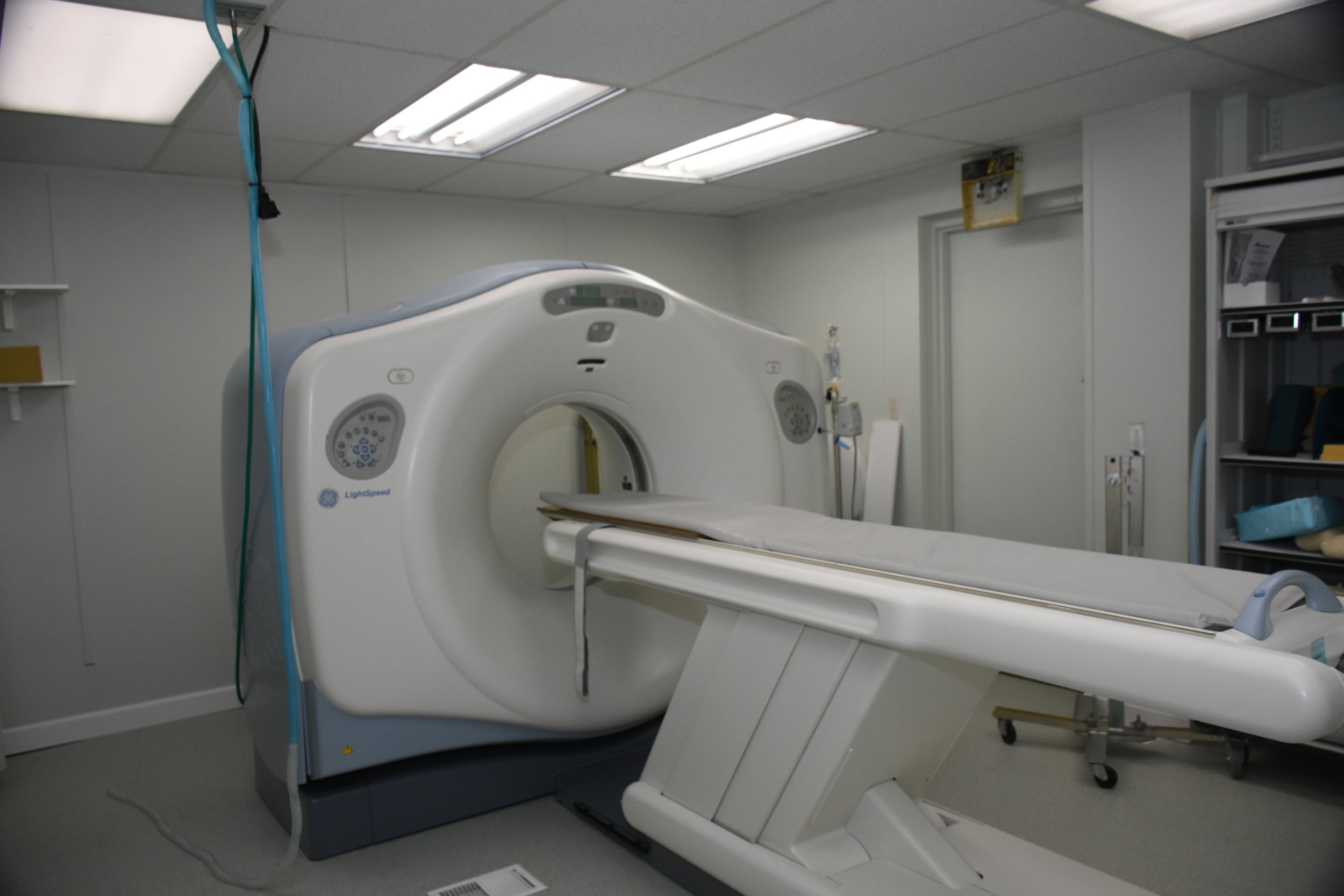
We are pleased to offer in-house CT scanning. The advanced computer systems in our new GE 16-slice helical scanner allow for scans to be completed in less than 3 minutes in most cases. CT has much greater resolution than plain radiography or ultrasound and therefore, is an important tool for diagnosing and staging cancer. We are often able to detect disease on CT that is not visible on ultrasound or plain radiographs, thereby improving our diagnostic accuracy.

CT scan of the lungs of a dog. The arrow points to a tumor in the lungs that was not visible on plain X-rays
A plain radiograph (X-ray) was taken and a large primary lung tumor was seen. On CT, however, a second tumor was found that was not clearly visualized on plain X-rays. Therefore, our treatment course was changed based on this CT. CT scans are used for a wide variety of applications.
Brain tumors, for example, do not show up on routine radiographs, because the skull blocks our ability to visualize brain structures on routine radiographs but are seen on CT.

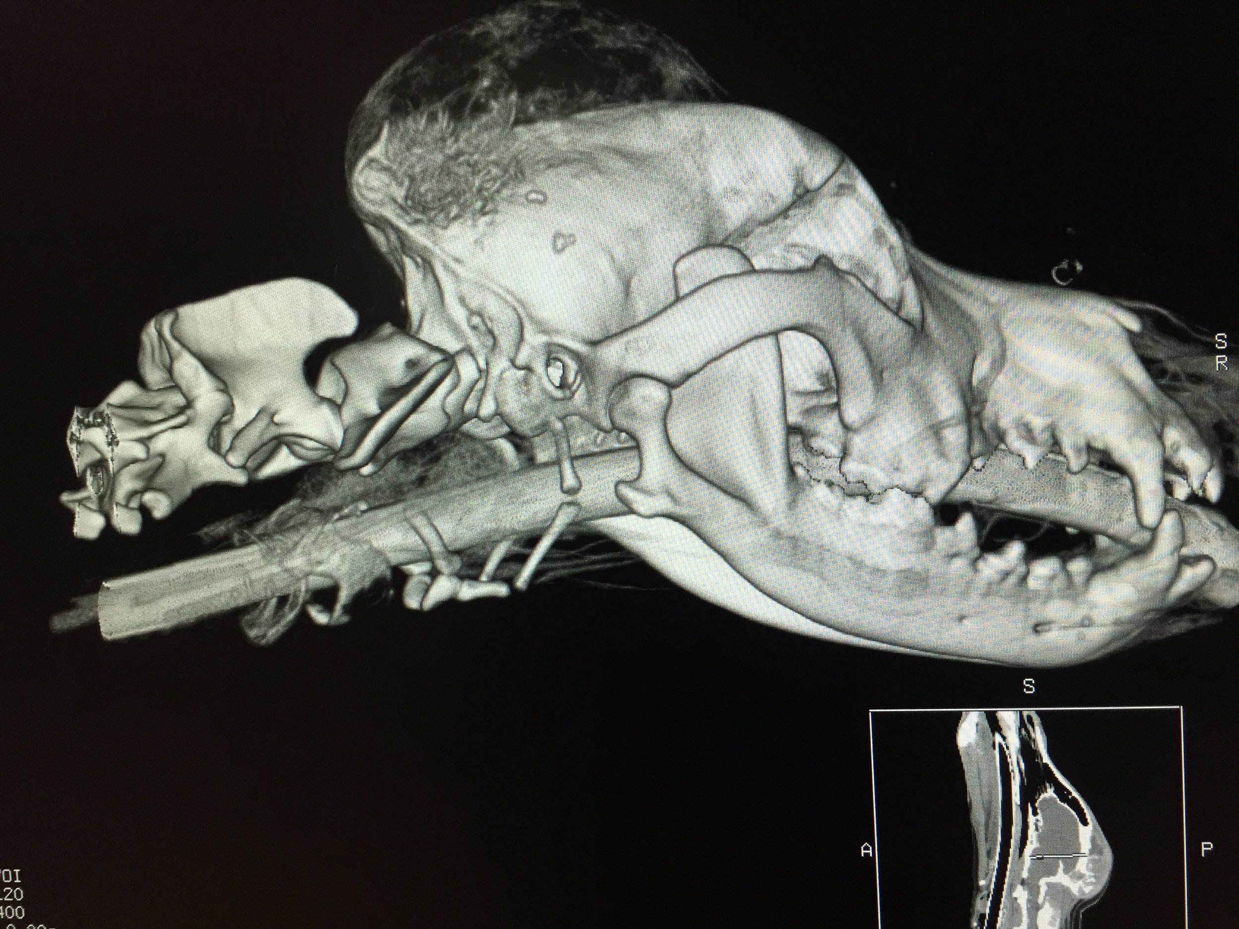
The nasal cavity is a very intricate region and excellent detail is needed to accurately evaluate patients for nasal disease. This is the scan of a Labrador retriever showing a normal nasal cavity. The intricate scroll-like structures seen in the center of the scan are the turbinate bones.
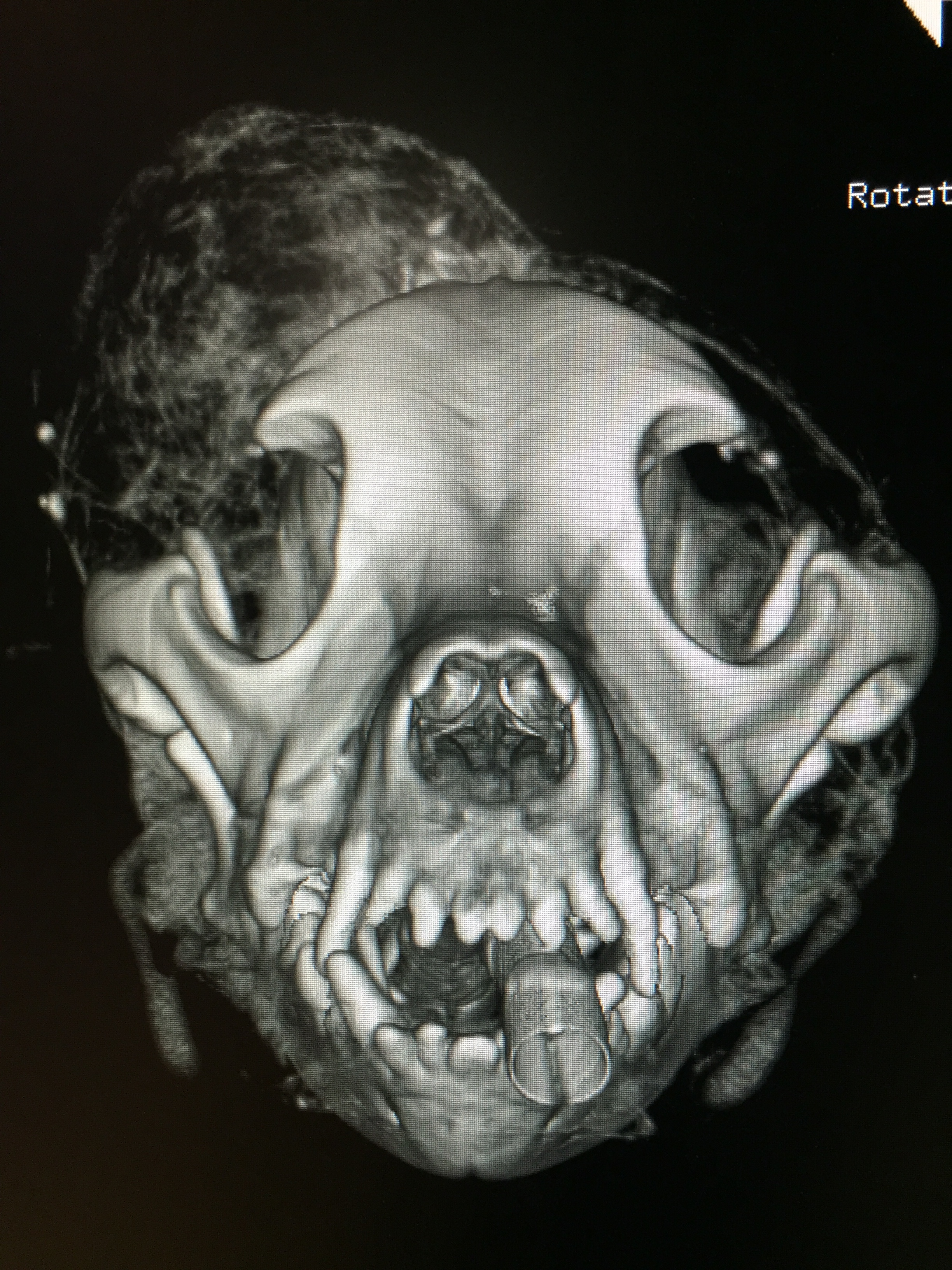
Bone reconstruction of a CT scan of the head showing a large mass on the forehead. The reconstructed images give us a more accurate and detailed picture of a tumor to help in surgical or radiation planning.

Contrast CT image of the brain of a cat. The arrow points to a large tumor that had the characteristics of a pituitary macroadenoma. This tumor was treated with radiation therapy. You can see an 'after' of Max at the end of this page!
How is a CT scan done? CT scans are performed while patients are under general anesthesia. Therefore, prior to a scan, blood work is required to be sure the patient is healthy enough to tolerate general anesthesia. In patients suspected of having cancer, multiple chest radiographs are also required prior to anesthesia. Patients are usually given an injection to induce or start anesthesia, and maintained on gas anesthesia until recovery.
Once under anesthesia, patients are positioned on the scanner. The images are taken by the CT scanner and processed by the computer to give us the information we need. It is usually necessary to give an IV “contrast” agent to help us image certain structures more precisely. For example, the image of the brain tumor shown on the previous page is with contrast. It brightens the image of the tumor aiding in identification.
What are some uses for CT in animals? There are many instances in veterinary medicine where CT aids in the diagnosis of tumors and other diseases and helps us determine the extent of disease present. The use of CT is especially helpful in diagnosing brain and nasal tumors, as these are difficult to impossible to see using plain radiography. If radiation therapy is used to treat brain or nasal tumors, a CT is required in order to generate a radiation treatment plan. Without CT, we would be unsure of the exact location and size of these tumors. In the case of a suspected primary lung tumor, CT is extremely helpful in determining the exact location of the mass and if there is any more disease present in the lungs that may not be visible with plain radiography. The CT also helps us determine if the lung mass is operable before we make an attempt at surgery.

The CT scan performed on this patient was converted to a 3-dimensional computer based treatment plan for radiation therapy. The use of CT based planning increases the accuracy of radiation delivery and decreases exposure to surrounding normal tissues.
INTERPRETATION of ct scans
The doctors at ACIC will preliminarily evaluate the CT scans at the time they are performed. All scans are then electronically sent to Dr. Sheila Etue, a board certified radiologist, for more in-depth evaluation. Reports from Dr. Etue are usually available within 1-4 days of submission. STAT reporting is also available.

This is a CT scan of a patient diagnosed with osteosarcoma in the front leg. Several years after treatment, the patient became painful in the hind limbs. Routine radiographs were normal, so a CT scan was recommended. The red arrows point to a lesion in the spine that represented spread of the osteosarcoma. This patient underwent palliative radiation.

This CT scan is of the abdominal cavity of a cat. The measured mass is an adrenal tumor. With CT we were able to determine that the mass was adrenal in origin and that there was no other evidence of disease, making the patient a good candidate for surgery.

Maxwell Behringer was diagnosed via CT scan with a pituitary macroadenoma.
His CT is the cat brain mass from above. He underwent a full course of radiation therapy in 2002 and lived a full and normal life for many more years.