
What are Nasal Tumors in Pets?
NAsal Cancer
Nasal tumors comprise approximately 2% of all cancers seen in dogs. Approximately two-thirds of all canine nasal tumors are carcinomas , including adenocarcinoma, squamous cell carcinoma and undifferentiated carcinoma; and one-third sarcomas , including fibrosarcoma, chondrosarcoma, osteosarcoma, and undifferentiated sarcoma. Other types of malignancies, including lymphoma, are possible but rare. Medium and large breed dogs appear to be predisposed. Although unproven, it has been suggested that long nosed breeds, dogs living in urban environments, and dogs exposed to tobacco smoke may be at higher risk. Nasal carcinomas are locally aggressive tumors with a low to moderate metastatic rate. When metastasis occurs, spread is typically to the lungs or regional lymph nodes. Tumor extension into the front part of the brain may also occur.
What are the symptoms of nasal tumors?
The most common symptoms include a nasal discharge which initially is unilateral and can be bloody (epistaxis) or mucopurulent or both. At times, epistaxis can be severe and may be difficult to stop. As the tumor grows, facial deformity from bone erosion of the tumor and a discharge from the eye as a result of obstruction of the nasolacrimal duct are seen. Other conditions such as fungal disease, bacterial or fungal rhinitis, or nasal foreign bodies can present similarly.
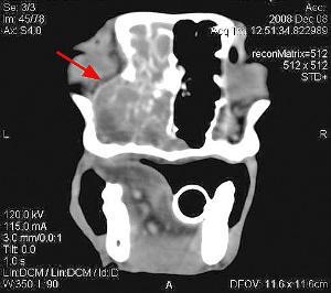
CT has superior image quality over conventional radiographs and allows us to see in great detail the extent of tumor.
How is the diagnosis made?
A presumptive diagnosis is made based on clinical symptoms, physical examination, and plain radiography of the nasal cavity. However, a definitive diagnosis is made through a tissue biopsy. Prior to biopsy, blood tests are done to confirm that the patient’s clotting ability is normal. Ideally, a CT scan (computed tomography) is performed immediately prior to the biopsy being taken.
Staging the disease:
Once a tentative diagnosis is made, and prior to advanced imaging, we recommend that basic staging tests be performed.
A complete clinical work-up includes:
Complete blood count (CBC), serum chemistry panel, urinalysis, and clotting profile.Thoracic radiographs (3 views)If blood work and radiographs are normal, proceed with CT to determine localized extent of disease with biopsy to follow. Because nasal tumors often start in the back portion of the nasal cavity, extension into the brain is possible. CT can determine if this has occurred.
Treatment and prognosis for nasal tumors:
Without treatment, the median survival for patients with nasal carcinomas is 95 days. Patients with some tumor types such as chondrosarcoma, can survive longer periods without treatment, but the majority of patients with other tumor types show relatively rapid progression of disease.

RADIATION THERAPY:
The treatment of choice for most nasal tumors is radiation therapy. Radiation therapy is delivered daily, Monday through Friday, over a 3-4 week period of time. Reported median survival times range from 8-19 months. With computerized treatment planning using CT (standard of care at ACIC) median survival times are improved due to the ability to greatly enhance dose distribution to the tumor while sparing normal tissue.
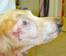
A severe example of desquamation after radiation therapy in a dog treated for an extensive nasal carcinoma. Complete healing occurred in approximately 3 weeks. The reddened area near the eye was caused by the patient rubbing his face on the carpet while the tender skin was healing.
RADIATION FOLLOWED BY SURGERY:
When surgery is performed on any residual tumor tissue left after radiation therapy, a median survival of 47 months compared to 19 months with radiation alone was recently reported. (J Am Vet Med Assoc 277:936-41, 2005). However, the combination treatment was associated with an increased incidence of serious late side effects. Larger studies are needed to fully evaluate this combination of therapy.
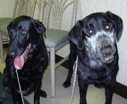
The Black Labrador on the right is shown with a new “mask” of white hair following radiation therap
ALLIATION WITH CHEMOTHERAPY:
The addition of chemotherapy to radiation therapy has not resulted in significantly improved survival times. It is, however, often effective in relieving clinical signs. Median survivals of 5 months are reported for patients with nasal adenocarcinoma treated with cisplatin chemotherapy (J Am Vet Med Assoc 200[3]:355-357 Feb 1’92). A more recent study presented at the Veterinary Cancer Society meeting in 2003 showed survival ranges of 5-32 months when doxorubicin, carboplatin and piroxicam were used in combination.
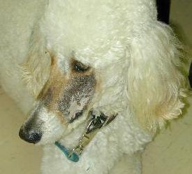
The Standard Poodle is shown with his new brown hair
PALLIATION WITH PIROXICAM ALONE:
Piroxicam is a non-steroidal anti-inflammatory drug(NSAID) that has been shown to have anti-cancer benefit for some tumors including canine carcinomas. This drug can potentially temporarily improve clinical symptoms in approximately 60% of patients treated.
What are the side effects of radiation?
Side effects of therapy can include hair loss in the radiation field (hair will usually grow back a different color), sun-burn like reaction to the skin in the radiation field, inflammation of the tissue in the oral cavity, and dry eye if the eye is in the radiation field (blindness is rare but cataracts are common). Side effects are typically moderate, but can in some cases be significant. However, even in more severe cases, healing is usually rapid.

Pre-radiation therapy CT scan. Red arrow points to tumor.

Same patient post-radiation therapy. CT scan shows resolution of tumor!